Archaeologists and hospital staff in England have collaborated to CT scan the head of an ancient Egyptian mummy discovered in an English attic.
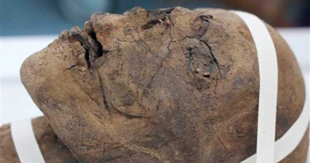
In the 1820s, a 2,700-year-old Egyptian mummy was sent as a souvenir to an individual in Ramsgate, a seaside town in the district of Thanet in East Kent, England. Following the death of the homeowner, his brother donated the head, housed in its glass display case, to the Canterbury Museums and Galleries collection, as reported by Kent Online.
Charting Ancient Corpes
Craig Bowen, the galleries collections and learning manager at Canterbury Museums, stated to the press that the mummy’s head was “discovered by a man who inherited it from his brother, who received it from a ‘Dr. Coates’ sometime in the early/mid-twentieth century.”
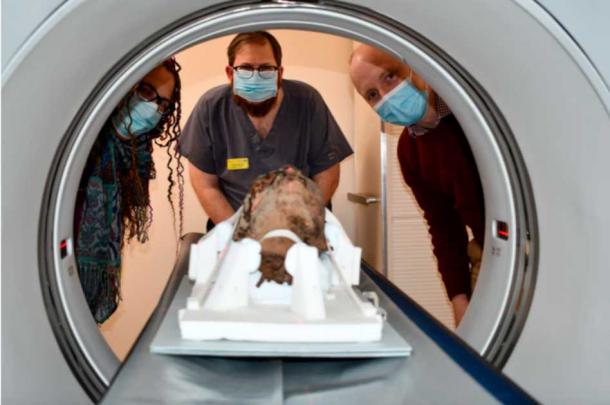
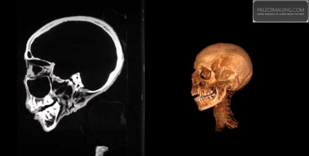
The X-rays conducted at Canterbury Christ Church University in 2020 determined that the mummy was an adult female. However, a comprehensive CT scan has now uncovered a wealth of new information about the individual before her death. CT scans, or ‘computerized tomography,’ entail capturing a series of X-ray images from various angles around a body. A supercomputer then integrates the cross-sectional images (slices), generating a high-resolution map of the scanned body.
Brainless in the Afterlife
The new CT scans were conducted by James Elliott, a lecturer in diagnostic radiography at Canterbury Christ Church University and senior radiographer at Maidstone and Tunbridge Wells NHS Trust. In a report on his website, Paleoimaging.com, Elliott states that the new scans offer “a significant amount of information, covering everything from dental status, pathologies, method of preservation, as well as aiding our estimations of age.”
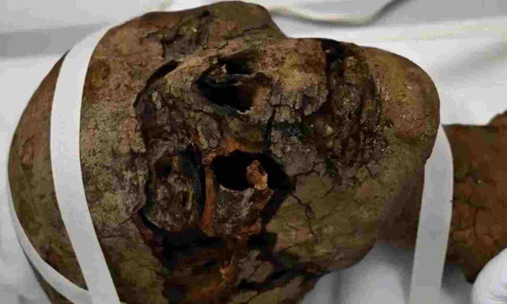
The scans revealed the removal of the woman’s brain, indicating that she had undergone a process at a Per-Nefer, or ‘house of beauty,’ after death. This location served as the initial stage for purification and mummification procedures and rituals. Traditionally, embalmers utilized hammers and chisels to access the brain through the nasal bone, inserting an iron hook to gradually extract the brain matter. The remains were then scooped out with a spoon, and the cranial cavity was rinsed with water.
Craig Bowen noted the irony that ancient Egyptians believed a person’s mind “was held in their heart and had little regard for the brain.” He also mentioned that CT scans are revealing “significant variability” in the methods of brain removal.
Details of the Ancient Egyptian Uncovered Through Mummy Head Scan
The CT scans revealed that the woman’s tongue was very well preserved, and her teeth showed signs of significant wear. The former attests to the advanced methodology of the ancient preservers, while the latter suggests that the woman had a lifelong diet of coarse foods.
According to a report on Kent Online, an unidentified material was discovered trapped within the mummy’s left nostril, described as “a tubing of unknown material.” The same substance was also identified in the spinal canal. The origin and composition of these blockages are currently unknown, and they could potentially be modern, possibly from the Victorian era.
Craig Bowen mentioned that this project is part of a broader effort to preserve the head and enable its display in conservation-grade packaging for public viewing. The team intends to utilize the newly obtained CT scanning data to produce a three-dimensional replica of the woman’s head. Additionally, they plan to reconstruct the woman’s face in 3D “without exposing the actual artifact,” as stated by Bowen.